 The year's best pictures from the world of medicine
[ 2009-10-21 11:41 ]
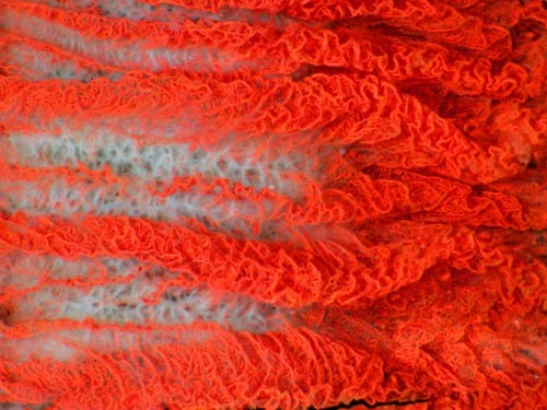
4 公牛眼睛中的毛细血管
这张光学显微照片是由斯匹克-沃克尔(Spike Walker)拍摄的。照片拍摄的是一只公牛眼睛睫状体的毛细血管。这些毛细血管能分泌水状液。这些液体为眼球晶体和角膜提供了大部分营养成份。
这张图片是从不同深度拍摄的27张照片合成而得到的,给人以三维图的效果。为了更突出显示公牛眼睛睫状体的毛细血管并更好地进行拍摄,毛细血管中注射了一种不可溶的染料。

This light microscope image by Spike Walker is of blood capillaries in the ciliary body of an ox's eye: the tiny holes that secrete a liquid called aqueous humour are shown. This liquid provides most of the nutrients for the lens and cornea. This image was created from a stack of 27 images taken at different depths and combined to give a three-dimensional appearance. The capillaries have been made visible by injecting an insoluble dye into the artery. (Image: M. I. Walker) [NewScientist]
|