 Best pictures from the world of medicine
[ 2009-10-21 14:04 ]
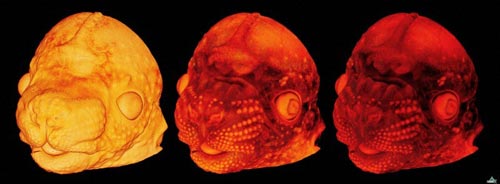
12 早期胚胎发展阶段的老鼠头部
这张3D图片显示的是早期胚胎发展阶段的老鼠头部,是由高清晰反相显微镜拍摄的。在拍摄过程中,样本放在塑料片上,然后涂上曙红荧光色。这种显微镜薄片切片机可切割非常薄的样本,最薄达到2微米。
使用计算机软件,老鼠头部的不同结构得以成像。图片是由英国医学研究理事会所提供的。

This 3D image of a developing mouse head at an early embryonic stage was created using high-resolution episcopic microscopy (HREM). In HREM, samples are embedded in a plastic that has been stained with eosin, a fluorescent dye. A microtome is used to slice away fine sections of the sample – as thin as 2 microns – then an image of the remaining block is captured after each slice is taken. Using computer software, different structures within the head can be visualised. Colour channels can be added to the 3D reconstruction to highlight tissue density, as shown here. (Image: NIMR, MRC) [NewScientist]
|